In this audio-visual essay, we bring the act of looking and listening to the foreground. We first invite the audience to observe the images and listen to the audio, and participate in the diagnostic puzzle of thinking about how these elements might connect. In the subsequent written essay, we unfold the puzzle slightly and raise more questions than answers to the themes of the objectivity of images and the science of the general versus the science of the particular. Finally, we discuss the space of the hospital and the museum to comment on the online format of viewing bodies.
Key Words human body, visualisations, spaces, museums, stakeholder collaborations
PDF
Looking at Bodies: In, At, Up, and Under
DOI: https://doi.org/10.51002/trajectoria_022_07
(Published March 31, 2022)
(Published March 31, 2022)
Abstract
Element 1
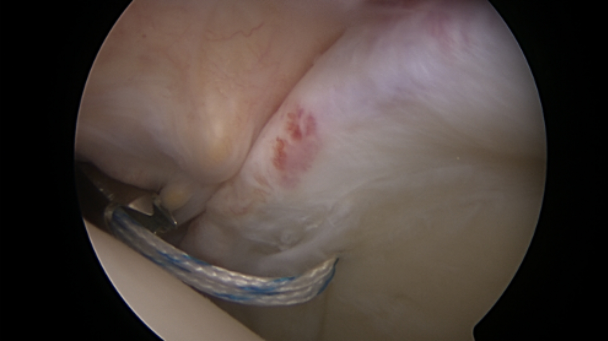 Figure 1 Medical Image from a Keyhole Surgery (Arthroscopy). The image shows the inside of the shoulder joint of an orthopaedic patient.
(©Medical Museion)
Element 2
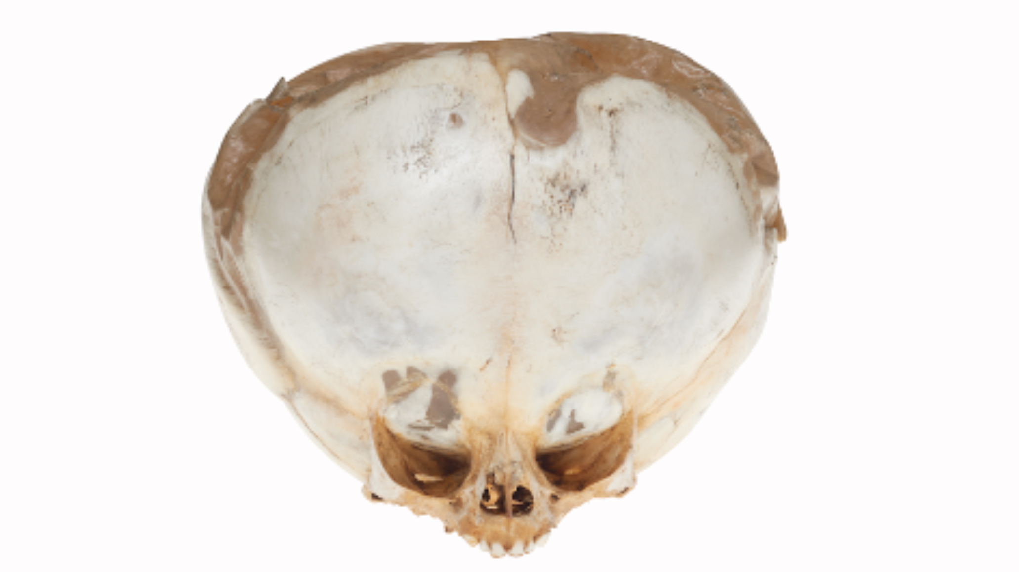 Figure 2 Contemporary Art Photograph of a Specimen from the Nineteenth Century: The Skull of a Child Who Suffered from Hydrocephalus
(©Nicolai Howalt, Medical Museion)
Element 3
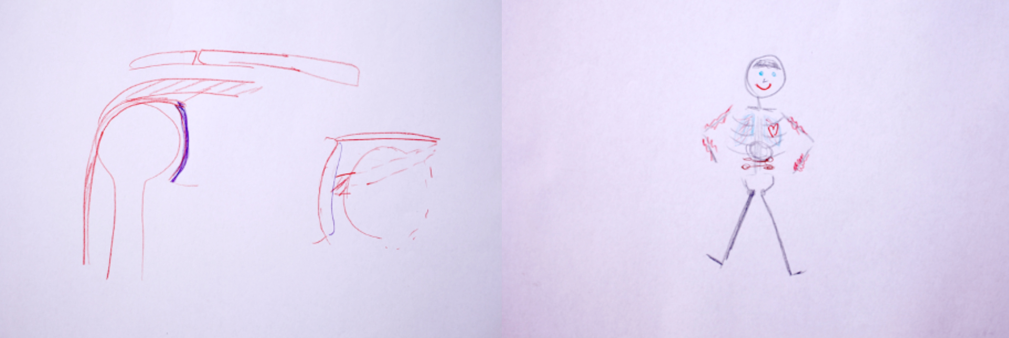 Figure 3 Pencil drawings by the patient whose shoulder image is depicted in Element 1
(©Medical Museion)
Element 4
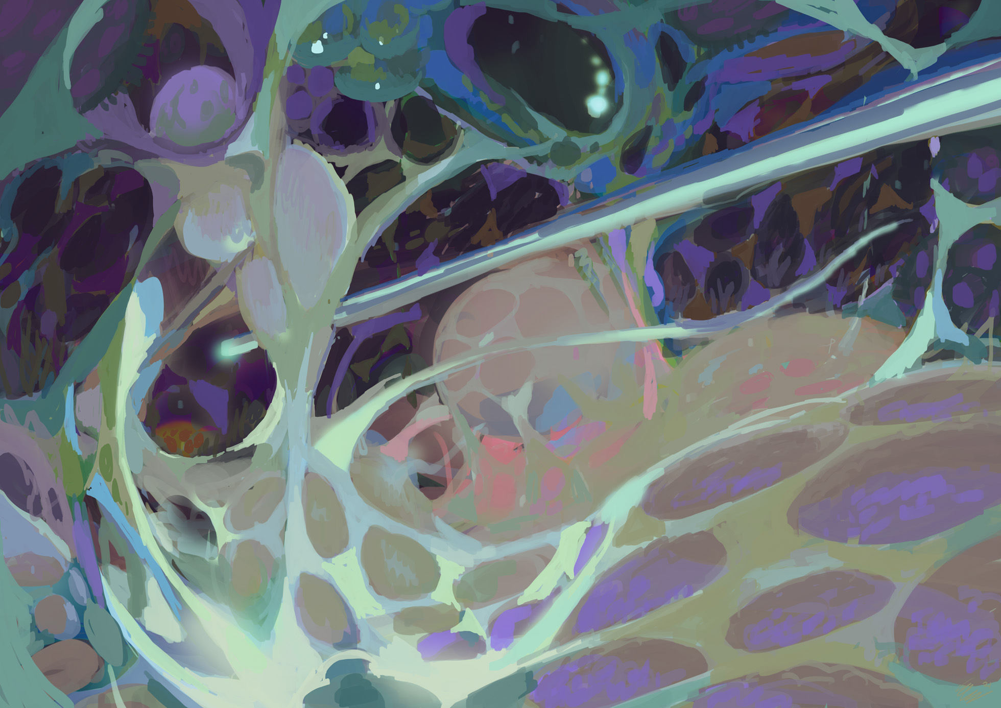 Figure 4 Illustration by Emil Friis Ernst of the Inside of the Patient’s Shoulder, Created Uniquely for This Submission
(©Emil Friis Ernst, Medical Museion)
Element 5
A Voice-over (Audio) with the Following Quotes:
Patient: I remember how surprised I was at how clean and neat it was in there. And also the colours. They were pretty colours. I do not know why I had imagined that there would be blood or – I just remember how fascinated I was at how clean and neat it was in there. I remember how they used this water to rinse, so they could see – and then you could see these small cilia. I think it just looked like a universe somehow.
Patient: Like outer space or under water. With corals. This thing with the nice colours and cilia or fibres. I think it is fascinating. And then I remember how I thought this claw that he used to catch the wire, I saw it as incredibly large. So he showed me this tool afterwards, and it was almost just the size of a sowing needle – I thought “Oh boy”
Patient: But the colours are pretty. And the shapes – things are rounded. I am also fascinated by the veins. It does not say a lot, but it almost looks like cod roe before it goes into the pot.
Patient: I am not disgusted by these things, or think they are disgusting or scary. I think it is fascinating. This skull where it says: Child, 6 months, water on the brain. When I see it I think of an alien. It is probably the image today’s world have created of an alien. We have not seen an alien, but the film industry has created this look: The very large head, the small mouth and eyes. I do not know about the shoulder, if it was mainly like outer space, or under water.
Patient: I am thinking, it is a peaceful and quiet place. Perhaps it is a bit difficult to explain. It is an unproblematic place—there is just…calm. It is not as if a lot is happening. If you imagine viewing inside a heart—I imagine—it would just be pumping away. That would be different from looking inside something that is just dead.
Patient: When I say dead, I mean completely quiet—as if a probe is sent down to a depth of 2–4 km, and it rushes over the ocean bed. Then nothing happens aside for stuff that is whirled up. It is not completely dead because of life and motion. It is similar to entering another universe inside the shoulder that we have not seen before.
(June 2021; Medical Museion, Copenhagen; Interviewer: Simone Grytter)3)
Reflections
1
Come look. Observe closely. Watch the fleshy, soft shapes of vague salmon-pink tissue laced up with a blue and white wire; look at the abnormal skull; the simple pencil drawing of blue, red, brown, and black; and the illustration of something otherworldly. If you listen, you can hear a voice talking about outer space and the deep sea. Do you have an idea of what you are looking at and how these elements might connect?
2
You are looking at images that are tied in different ways to one person. They have all been experienced, looked at, talked about, invented, or created by the same person. Some might call them representations, renderings, or mediations; others might call them presentations, reality, or raw material. Regardless, they all draw a line back to the same body, the same individual, patient, informant, museum visitor, and human being. The elements are connected and unfolded from within the singular individual.
3
This project has its home in Medical Museion (https://www.museion.ku.dk/en/), a contemporary medical museum, research group, and a section of the Public Health Department at the University of Copenhagen.4) Here, explorations of elements inside human beings have historically focused on specimens of the deceased. Specimens such as organs, skulls, or entire bodies were collected by doctors to investigate pathologies and teach other doctors; these specimens were later donated to the museum. However, in this project, we leave the museum and enter the hospital to explore current ways of peeking inside the living body. The project builds on ethnographic fieldwork at an elective orthopaedic surgery unit in a large hospital in Denmark. Here, Grytter observed keyhole surgeries performed on the shoulders, knees, and hands, where patients are awake, and watch the procedure on a screen. After the surgery, patients were interviewed about their experiences of following their surgery on screen. A few patients were also invited to Medical Museion, more than a year after their surgery, to explore what happened when they watched other bodies and specimens in a museum context. During this session, the patients also drew their own bodies, viewed exhibitions at the museum, and discussed watching all types of bodies. We engaged with one of these patients for this exploratory investigation, where we bring together and allow touching and smearing of theoretical surfaces and methodologies in the research fields of medical humanities, visual anthropology, and curatorial practices. Using different research perspectives, we aim to probe the understanding of the anatomy of one person and the (re)presentations that might permeate and emerge from this singularity.
4
We delve deeper into the elements presented in this study by first looking inside the body, utilising an image created in a physical space that is traditionally used to look inside, examine, and treat the human body—a hospital. More specifically, we are looking at a medical image of the inside of an orthopaedic surgical patient’s shoulder (Element 1). The image was created during arthroscopy, a keyhole surgery, where a small camera is inserted in the patient’s shoulder through a small incision. The camera becomes the eye of the surgeon, and the camera’s view is projected on a large screen in the surgical theatre. We are swimming in the joint along with the surgeon (Prentice 2014). However, using the pronoun ‘we’ is not appropriate in this context. In the operating theatre, the patient can choose to be awake during the surgery, which provides them an opportunity to look inside their own body in real-time—a rare glimpse of the interior landscape of humans.
5
In the next element (Element 2), we explore a contemporary photograph by the artist Nicolai Howalt of a skull. The skull is exhibited in Medical Museion’s exhibition ‘The Body Collected’ (https://www.museion.ku.dk/en/bodycollected). This element is an artistic interpretation—or rendering—of a specimen from the nineteenth century and illustrates the skull of a young child who suffered from hydrocephalus (water on the brain). One may wonder why is this image included in this essay next to the image of the inside of a patient’s shoulder. We invited the patient who had experienced looking inside his own body on a screen to watch other bodies at Medical Museion. When observing the various human specimens at the museum, the patient paused by this skull. ‘This reminds me of an alien’, he said, and continued to compare what he saw inside his shoulder during the surgery to an imaginary world, looking up into outer space, and deep down under water. The audio you heard is an excerpt from this conversation.
6
In Element 3, we see a pencil drawing of a smiling stick human with red lips, blue eyes and lungs, and a red heart. The patient created this image when asked to draw on a piece of paper how he appeared from the inside. In the other drawing, the patient illustrated how he imagined his shoulder bone fits and moves around his shoulder. The patient drew these after watching how his shoulder appeared from the inside.
7
To push the process of image creation to the foreground, we decided to actively engage in the production of further images. We collaborated with the science fiction illustrator Emil Friis Ernst (https://www.beingernst.com) and asked him to draw the world that the patient described when he observed the skull in the museum and thought about how he experienced looking inside his body during the surgery. We did not ask the illustrator to draw something particular, nor did we provide directions on a specific choice of style or use of colours; rather, we offered him the flexibility to interpret the patient’s quote on his own. The image is not merely a commissioned illustration, but the expression of an artist’s creative imagination. A wide body of literature has already established how the understanding of the human body affects and is affected by its portrayals in popular cultures such as sci-fi films, cartoons, photography, and exhibitions. Kirsten Ostherr (2013) writes about how the roles of patients and doctors are continually shaped through media such as advertisements, television programmes, cinema, medical films, and imaging technologies. José van Dijck (2005) investigates how the understanding of the inner body is always created and mediated through technology, artistic interpretations, and social norms. Elizabeth Hallam (2016) writes about how anatomy is displayed in museums and states that anatomy is a field of knowledge that is never fixed but always ‘constituted and communicated in practice within particular social and cultural contexts’ (Hallam 2016: 8).
8
The abovementioned studies suggest that our understanding and experience of the human body has always been shaped and altered by various medical, technological, societal, and cultural influences. We also know how we engage actively in this dialectic loop by co-producing and integrating further visualisations of the body—created from and with the patient’s experience—into it, thereby providing a further imaginary body. Exploring and understanding the body within the cultural realm of science fiction has been conducted several times, and to further engage in the loop is perhaps verging on cliché. Why do we pursue the repetition of this science fictional realm of reference? The main reason lies in the patient’s words when he describes looking inside his own shoulder as if it were a universe filled with an alien, the outer space, the deep waters—narrative elements belonging to a universe of science fiction. Therefore, the interesting question here might be why the patient chose to describe the aesthetic of his own inner body through these metaphors of outer space and the depth of the oceans.
9
Other examples of science fiction being employed to envision the human body emblematically include the 1966 sci-fi film Fantastic Voyage, where a team of explorers is shrunk to a minute size to venture on a dramatic journey inside the body of an important scientist to cure a blood clot in his brain. Another example is the crumbling borders between the human body and machine, animal, nature, and other non-human elements manifested in the character of the cyborg, explored in the theoretical works of Donna Haraway. Haraway is inspired by the science fiction writer Ursula Le Guin and her carrier bag theory—a narrative style without a hero, without linear progress—that tells stories that might be messy, entangled, and conflicted (Le Guin 1989). Storytelling and how narratives are built become essential. ‘It matters what stories we tell to tell other stories’ (Haraway 2016: 118). What are the narratives we use to tell stories about bodily anatomy? At the beginning of this essay, we introduced the opportunity to tell stories of the human body through what can be called a nonlinear method, or the method of collage, montage: the distributed. Borrowing from Annemarie Mol when she said that ‘reality is distributed’ (Mol 2002: 96), the body is not just understood in one singular way but is created based on a multiplicity of practices and contexts—in the hospital, museum space, and this online journal format. Depending on their juxtaposition, in this article, we suggest multiple rather than just one understanding of particular human anatomy.
10
The incorporation of art and the artistic workings of a given phenomenon is, of course, very familiar in the world of museums and galleries, where curators increasingly often mix exhibits (evidence) that originate in the different worlds of art and science. This is frequently implemented at Medical Museion, where maintaining an artistic eye on the scientific field has been the key in exhibitions such as Mind the Gut (https://www.museion.ku.dk/en/mindthegut/), and The World is in You (https://www.museion.ku.dk/en/the-world-is-in-you/). The use of art can be accepted more readily by audiences than by discipline-based commentators and critics, and artistic practice does not play a role in, for instance, the surgical space. In this science fiction illustration, we wish to melt together patient experiences from the medical and the museum spaces, step out of our role as observer-researchers, and actively facilitate the production of new images.
11
Thus, given that the origin of the elements in this essay, how they emerged, and how they connect to the same person is now known, we can consider how close to reality the different elements are, and whether one is closer than the others to the status of being a raw material. Perhaps the patient’s own account should be of special significance, as the body belongs to him after all. What about the drawing—can it be considered less important because the patient could be more competent at describing than drawing, or because the medical image could better reflect the truth than the pencil drawing of a stick human? This may be true only if one has sufficient medical training to know and understand what they are looking at. Images, especially those produced in a medical context, may be read or viewed as rendering the body transparent and as carriers of scientific facts (see Holtzmann Kevles 1997), and we tend to understand images of anatomy as actually watching the true interior of the body (Stephens 2011: 3). Others have argued that the body will never become transparent and that medical images constitute another layer or representation of a body that we can never fully understand (van Dijck 2005). What is the power relation between the images—are some of them more truthful than others? Are the elements presented in this essay simply different types of representations of a body?
12
Lorraine Daston (2014: 320) notes that ‘the time is ripe to think about images beyond representation’, and argues that a representation naturally presumes a presentation, implying something that occurs before the remaking, something original that has an ontological status. If images are to become more than mere representations of something else, we need to blend the presentation and the representation, the subject and the object (Daston 2014). On the one hand, the images at the beginning of this essay might be viewed as different representations or renderings of the patient. On the other hand, the elements can be viewed as no longer being attached to their origin, and thus, not representing the patient—in this online forum, they might become free agents, to be understood independently. When approaching the images and audio pieces as not only representations of a patient’s body but also as presentations in themselves, we, in theoretical terms, view them as possessing agency (Gell 1998) and the ability to talk (Daston 2004), create new meanings, and affect and be affected by the praxis and context of which they are a part (Mol 2002). The interesting point here is not a discussion on whether the elements have agency but a focus on how the material elements act and create meaning within the specific spatial context of this online journal. Following Nicholas Thomas (1991), one might ask whether re-contextualisation implies ‘muting’ things. Rather, we view the elements as not merely representations of the patient but as having the ability to create new meaning in the online universe of TRAJECTORIA. The point is simple: for instance, the medical image of the patient’s shoulder adds to the creation of a specific understanding of the shoulder in the hospital context but reflects something different when curated for this online publication.
13
This essay has several spaces at play: first, the spaces of the hospital and the museum, although neither of these spaces is visible. The scenery of the surgical room or the skull placed in an exhibition case has no contextual images—we only observe the isolated objects that the patient was looking at. Spaces comprise much more than the act of looking or listening; they include heavily loaded atmospheres and sensory experiences. An orthopaedic surgical space has a very different atmosphere than the space of the museum; however, in both places, the atmosphere naturally affects the experience of what we observe. From where is this body of elements being watched and this text being read? It might be from a hospital or a medical museum in Denmark or anywhere else—from a house, on the bus, or from the office. This essay is most likely being read on the screens of a computer, tablet, or phone. We observe bodies in the hospital, museum, and online. However, in this online space, the atmosphere becomes a part of the meaning-constituting context very differently. We do not hear the drilling sounds and sense the sterility of the orthopaedic theatre, nor do we slide through the museum, glancing at other guests while inhaling the meaning and materiality of specimens or artworks.
14
We might ask if looking at bodies in this online space juxtaposes or collapses the walls between the museum, the hospital, and the viewer. In this online submission, we aim to create a space where we do not discuss only elements originating in a hospital or museum space, but also harness its supposed shortcomings—its lack of physical material context, absence of smell and touch, and its contextualised atmosphere. The online space becomes free-floating, where visual elements break loose from their original physical space and meaning and are only experienced through the flatness of the screen. We also do not know much about the patient from the elements presented to us and attributes such as age, occupation, and life, and feelings remain a mystery. By inducing this limitation, we intentionally reduce the patient to both a scientific as well as a museum object. The patient becomes an image that is presented here. Perhaps this goal of complete anonymity compels the visual elements to break free from their origin, the object retreats from the subject, and the representation from the presentation. The only element that informs the reader of the connection between the patient and the visual and audio elements in this project is this written part. What would happen if we simply removed this text and let the images speak for themselves? Which connections between the elements would then be formed? While working on this essay, we also considered how to draw the reader to stay with the visual elements in the beginning, how to prevent them from simply glancing over the images and jumping straight to these contextual words, and how to ensure that they treat the images with the same time, gravity, and ability to create knowledge as these words.
15
As researchers investigating medical practices and materiality, we are prone to acknowledge the science of the general. We analyse ‘x’ number of informants to provide sufficient data to assert something useful and validated about a medical practice. Nevertheless, in this piece, we engage with the science of a particular entity as we only cast our glance at one individual. ‘Conjectural knowledge’ is the term Italian historian Carlo Ginzburg (1980) applies to a type of understanding where particulars rather than generalities are thrust to the fore. He contends that its production is shared across methodologies, which, for example, art historians, psychoanalysts, and forensic detectives apply differently. Each of them pursues a form of ‘diagnosis’ guided by deciphering and amassing clues, held together by the ‘elastic rigour’ of ‘a whiff, a glance, an intuition’ (Ginzburg 1980: 28). Here, we apply this method of the diagnostic puzzle to encounter, uncover, and unfold a particular body. We also establish a riddle without one true objective solution and propose several different subjective experiences of a particular body—the patient’s experience, artistic expressions, our own curation, and the experience of the reader. As Annemarie Mol (2002) pointed out, even the particular body becomes multiple when it is performed or enacted in different contexts and relations.
16
In our experiment, our objective is not to offer the reader a deeper insight into how the human body is anatomically constructed; rather, we hope to provide a methodological quest—a focus on the process of how we search for connections between elements that are out of their original context. If the reader has been confused while observing the elements at the beginning of this essay—and potentially while reading this essay—then we have succeeded in our goal, especially if that confusion has been enlightening. Our aim is for the reader to frantically search for their connections and solutions. This process—to be searching without receiving one answer, especially while contemplating just one case—is what we intend the reader to absorb. We seek to trigger thoughts on how images emerge, how we understand them, and what their value and power are. We also intend to underline the epistemological value of engaging with the particular through fragmented experiences, and conversely, the understanding of the fragmented experience afforded by dwelling on the particular.
Notes
- 1)
- The author, Simone Grytter, is a PhD student at Medical Museion and Centre for Medical Science and Technology Studies.
- 2)
- The author, Ken Arnold, is director of Medical Museion, part of the Novo Nordisk Foundation Center for Basic Metabolic Research.
- 3)
- This conversation has been reenacted by Adam Bencard, respecting the informant’s right to anonymity.
- 4)
- This essay is a part of a Simone Grytter’s PhD project that investigates what happens when we view inside our own living body, and how this might affect the way we experience it.
References
- Daston, L.
- 2004
- Introduction: Speechless. In L. Daston (ed.) Things That Talk: Object Lessons from Art and Science, pp. 9–26. New York: Zone Books.
- 2014
- Beyond Representation. In C. Coopmans, J. Vertesi, M.E. Lynch, and S. Woolgar. (eds.) Representation in Scientific Practice Revisited, pp. 319–323. Cambridge, Massachusetts: The MIT Press.
- Gell, A.
- 1998
- Art and Agency: An Anthropological Theory. Oxford: Clarendon Press.
- Ginzburg, C.
- 1980
- The Cheese and the Worms: The Cosmos of a Sixteenth-century Miller. London: Routledge & Kegan Paul.
- Haraway, D. J.
- 2016
- Staying with the Trouble: Making Kin in the Chthulucene. Durham and London: Duke University Press.
- Hallam, E.
- 2016
- Anatomy Museum: Death and the Body Displayed. London: Reaktion Books Ltd.
- Holtzmann Kevles, B.
- 1997
- Naked to the Bone: Medical Image in the Twentieth Century. New Brunswick, New Jersey: Rutgers University Press.
- Le Guin, U.
- 1989
- The Carrier Bag Theory of Fiction. In U. Le Guin (ed.) Dancing at the Edge of the World: Thoughts on Words, Women, Places, pp. 165–70. New York: Grove.
- Mol, A.
- 2002
- The Body Multiple: Ontology in Medical Practice. Durham and London: Duke University Press.
- Ostherr, K.
- 2013
- Medical Visions: Producing the Patient Through Film, Television, and Imaging Technologies. New York: Oxford University Press.
- Prentice, R.
- 2014
- Swimming in the Joint. In C. Coopmans, J. Vertesi, M. E. Lynch, and S. Woolgar (eds.) Representation in Scientific Practice Revisited. pp. 89–106. Cambridge, Massachusetts: The MIT Press.
- Stephens, E.
- 2011
- Anatomy as Spectacle: Public Exhibitions of the Body from 1700 to the Present. Liverpool: Liverpool University Press.
- Thomas, N.
- 1991
- Entangled Objects: Exchange, Material Culture, and Colonialism in the Pacific. Cambridge, Massachusetts: Harvard University Press.
- van Dijck, J.
- 2005
- The Transparent Body: A Cultural Analysis of Medical Imaging. Seattle: University of Washington Press.
Websites
Emil Friis Ernst
https://www.beingernst.com/ (Retrieved December 2, 2021)
Exhibition: Mind the Gut
https://www.museion.ku.dk/en/mindthegut/ (Retrieved December 2, 2021)
Exhibition: The World is in You
https://www.museion.ku.dk/en/the-world-is-in-you/ (Retrieved December 2, 2021)
Medical Museion
https://www.museion.ku.dk/en/ (Retrieved December 2, 2021)
Nicolai Howalt
The contemporary photography appears in the catalogue for The Body Collected
Medical Museion’s Exhibition: The Body Collected
https://www.museion.ku.dk/en/bodycollected (Retrieved December 2, 2021)